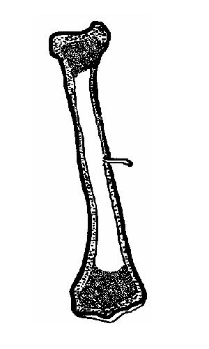
Long Bone Diagram No Labels : Do you know your funny bone from your femur?. Body anatomy organs human muscle anatomy human skeleton anatomy anatomy bones gross anatomy anatomy and physiology quiz muscular system anatomy muscle diagram anatomy coloring book. A long bone has two parts: There are many ways to approach the assessment of the radiograph; Labelled diagram of long bone. Blank muscle diagram to label.
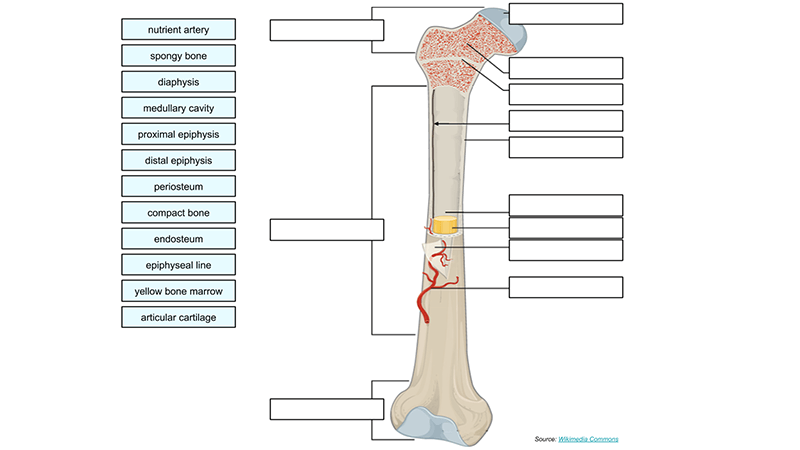
It is placed laterally to tibia and is the most slender of all the long bones. The covering of a bone. This is just one approach. There is a printable worksheet available for download here so you can take the quiz with pen and paper. In long bones, as you move from the outer cortical compact bone to the inner medullary cavity, the bone transitions to spongy bone.
6 2 Bone Classification Anatomy Physiology from open.oregonstate.education Describing a fracture is a basic requirement when making an assessment of a plain radiograph. The eight bones of the wrist are:. Anatomy and physiology of axial skeleton dinosaur skeleton human muscles skeleton diagram anatomical skeleton fish skeleton snake skeleton human spine anatomy models skeleton bones human skeleton. It contains the connecting cartilage enabling the bone to grow, and disappears at adulthood. In a long bone, for example, at about 6 to 8 weeks after conception, some of the mesenchymal cells differentiate into chondroblasts (cartilage cells) that form the hyaline cartilaginous skeletal precursor of the bones (figure 6.4.2a). This is just one approach. Circulatory system diagram circulatory system Long head (hamstrings) biceps femoris:
There is a printable worksheet available for download here so you can take the quiz with pen and paper.
Describing a fracture is a basic requirement when making an assessment of a plain radiograph. The spine contains 39 bones with the neck being quite long. The covering of a bone. Most, but not all, features you are required to know are shown on the following pages. All the above make up the circulatory system presented below: Medullary cavity cavity within the shaft of the long bones filled with bone marrow The interior part of the long bone is called the medullary cavity; Some bones are long and thick, like your thigh bones. A long bone has a shaft and 2 ends. Label skeleton diagram worksheet wiring diagram. Body anatomy organs human muscle anatomy human skeleton anatomy anatomy bones gross anatomy anatomy and physiology quiz muscular system anatomy muscle diagram anatomy coloring book. Labelled diagram of long bone. The eight bones of the wrist are:.
The red blood cells, white blood cells and platelets are formed by the bone marrow, which is a soft tissue in the bones. The bones of the hands can be divided into those that make up the upper arm, the lower arm, the wrist, the palm and the fingers. September 14, 2019 in worksheets. Metaphysis part of the bone between the epiphysis and the diaphysis; (b) in this micrograph of the osteon, you can see the.
Skeleton Worksheet Wikieducator from wikieducator.org It runs from the shoulder to the elbow. This bone is on the thumb side of the hand near the radius.; It is placed laterally to tibia and is the most slender of all the long bones. Legs, shoulder blades and ribs are examples of this type. 25 digestive system diagram no labels. One common treatment is rodding surgery, where a metal rod is inserted into a long bone to strengthen and prevent deformity. Humerus (2) radius (2) ulna (2) carpals (16) metacarpals (10) phalanges (28) total number of bones=60. It contains the connecting cartilage enabling the bone to grow, and disappears at adulthood.
The bones of the hands can be divided into those that make up the upper arm, the lower arm, the wrist, the palm and the fingers.
There are many ways to approach the assessment of the radiograph; Long head (hamstrings) biceps femoris: The neck and backbone of the chicken is very flexible. Spongy bone, also known as cancellous bone or trabecular bone, is a very porous type of bone. The eight bones of the wrist are:. Plasma is the liquid part of the blood, produced in the liver, which makes about half of the blood content. The covering of a bone. Newborn babies are actually born with many more bones than this (around 300), but many bones grow together, or fuse, as babies become older. The diaphysis and the epiphysis. The hollow region in the diaphysis is called the medullary cavity, which is filled with yellow. The red blood cells, white blood cells and platelets are formed by the bone marrow, which is a soft tissue in the bones. In the centers of these bones is bone marrow which makes blood cells. Bones of the axial and appendicular skeleton.
It runs from the shoulder to the elbow. Diagram of the digestive system. The tarsus or heel bone consist of 7 bones that make up the posterior part of the foot, that is present between the tibia, fibula and metatarsals. The interior part of the long bone is called the medullary cavity; This is an online quiz called label the long bone.
Label A Long Bone from www.biologycorner.com Circulatory system diagram circulatory system Bone · august 3, 2016. The outer shell of the long bone is compact bone, below which lies a deeper layer of cancellous bone (spongy bone), as shown in the following figure. The femur is the only bone located within the human thigh. A long bone has two parts: Do you know your funny bone from your femur? 25 digestive system diagram no labels. The calf bone or fibula is the smaller of the two bones that form the lower leg.
In the centers of these bones is bone marrow which makes blood cells.
Using what you know about the structure of a typical long bone, what part of the long bone is the most likely place for this. Touch device users, explore by touch or with swipe gestures. A dense fibrous membrane covering the surface of bones (except at their extremities) and serving as an attachment for tendons and muscles. Short bones are about as wide as they are long. The interior part of the long bone is called the medullary cavity; There is a printable worksheet available for download here so you can take the quiz with pen and paper. Enlarged terminal part of the bone, nearest the center of the body, made of spongy tissue and articulating with neighboring bones. A worksheet for labelling a long bone. Do you know your funny bone from your femur? Anatomy students in traditional classes may do practice labeling the bone on paper or even doing a coloring activity to help them learn the parts of the bone. The skull, humerus (arm bone), pelvis and collar bones are examples; Labelled diagram of long bone. This is a single long bone of the upper arm.
You need to get 100% to score the 10 points available long bone diagram. The inner core of the bone cavity is composed of marrow.


0 Komentar